User comb11doctor uploaded the image
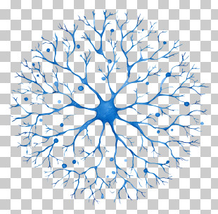
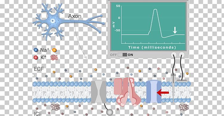
A digital illustration of a neuron cell. The neuron is shown in the top left corner of the image, with a blue and white color scheme. On the top right corner, there is a green screen with a graph that shows the time (milliseconds) of the neuron. The graph is divided into two sections. The top section shows the neuron's structure, with the neuron on the left side and the cell on the right side. The cell is made up of multiple layers of cells, each with a different color scheme - blue, red, and white. The cells are arranged in a grid-like pattern, with some overlapping each other. The blue cells are connected to the red cells, while the white cells are scattered throughout the image. The red cells have a red arrow pointing towards the neuron, indicating that the neuron is interacting with the cell in the center of the screen. The white cells appear to be interacting with each other, suggesting that they are interacting with other cells in a complex network. The image also shows a red and white arrow pointing downwards, indicating the direction of the time in milliseconds.
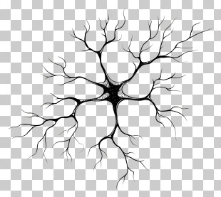
Diagram Neuron Resting Potential Action Potential Membrane Potential PNG
. The resolution of this PNG file is 1200 x 630 pixels and it has a file size of 387.01 KB.A digital illustration of a neuron cell. The neuron is shown in the top left corner of the image, with a blue and white color scheme. On the top right corner, there is a green screen with a graph that shows the time (milliseconds) of the neuron. The graph is divided into two sections. The top section shows the neuron's structure, with the neuron on the left side and the cell on the right side. The cell is made up of multiple layers of cells, each with a different color scheme - blue, red, and white. The cells are arranged in a grid-like pattern, with some overlapping each other. The blue cells are connected to the red cells, while the white cells are scattered throughout the image. The red cells have a red arrow pointing towards the neuron, indicating that the neuron is interacting with the cell in the center of the screen. The white cells appear to be interacting with each other, suggesting that they are interacting with other cells in a complex network. The image also shows a red and white arrow pointing downwards, indicating the direction of the time in milliseconds.
Diagram Neuron Resting Potential Action Potential Membrane Potential PNG
You might also like...