User carlsonart uploaded the image
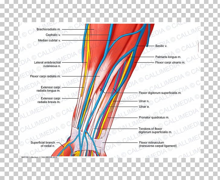
A cross-section of the leg, showing the muscles and ligaments of the foot. labeled with the names of the different types of muscles in the leg. The leg is shown in a red color, with the muscles highlighted in blue and yellow. The muscles are arranged in a radial pattern, with each muscle having a different color and shape. The ligaments are labeled with their corresponding names, such as "Lateral antebrachialis m", "Cephalic v", "Median cubital v", and "Palmaris longus m". There are also several other muscles visible in the image, including the flexor carpi radialis m, which is a type of nerve that extends from the foot to the ankle. These muscles are responsible for regulating the flow of blood and fluid in the foot, allowing for the movement of the bones and muscles to move freely. The flexor is responsible for providing support and stability to the foot and ankle, helping to reduce pain and discomfort.
Nerve Blood Vessel Forearm Muscle Vein PNG
. The resolution of this PNG file is 600 x 600 pixels and it has a file size of 360.89 KB.A cross-section of the leg, showing the muscles and ligaments of the foot. labeled with the names of the different types of muscles in the leg. The leg is shown in a red color, with the muscles highlighted in blue and yellow. The muscles are arranged in a radial pattern, with each muscle having a different color and shape. The ligaments are labeled with their corresponding names, such as "Lateral antebrachialis m", "Cephalic v", "Median cubital v", and "Palmaris longus m". There are also several other muscles visible in the image, including the flexor carpi radialis m, which is a type of nerve that extends from the foot to the ankle. These muscles are responsible for regulating the flow of blood and fluid in the foot, allowing for the movement of the bones and muscles to move freely. The flexor is responsible for providing support and stability to the foot and ankle, helping to reduce pain and discomfort.
You might also like...