User l3on uploaded the image
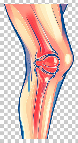
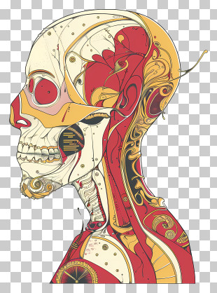
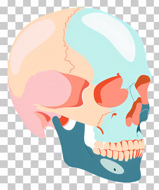
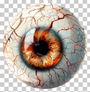
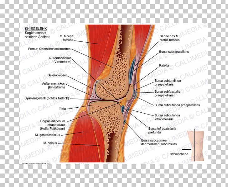
A cross-section of the knee joint, showing the bones and ligaments. labeled with the names of the different parts of the joint, including the knee, the femur, the bursa subpatella, and the tibia. The knee joint is shown in red and orange colors, with the bones in the center of the image. The bones are arranged in a radial pattern, with some bones on the left side and others on the right side. The femur is located in the top left corner, while the tibialis is located on the top right corner. The tibia is located at the top of the thigh, and it is located near the knee. The bursas subpatellaris are located on both sides of the femurs, and they are located near each other in the lower part of the body. The ligaments are labeled with their names, such as "Knee" and "Sagittalis", which are the names that make up the joint. Overall, the image shows a detailed view of the anatomy and physiology of the human knee joint.
Knee Sagittal Plane Human Anatomy Coronal Plane PNG
. The resolution of this PNG file is 600 x 600 pixels and it has a file size of 362.04 KB.A cross-section of the knee joint, showing the bones and ligaments. labeled with the names of the different parts of the joint, including the knee, the femur, the bursa subpatella, and the tibia. The knee joint is shown in red and orange colors, with the bones in the center of the image. The bones are arranged in a radial pattern, with some bones on the left side and others on the right side. The femur is located in the top left corner, while the tibialis is located on the top right corner. The tibia is located at the top of the thigh, and it is located near the knee. The bursas subpatellaris are located on both sides of the femurs, and they are located near each other in the lower part of the body. The ligaments are labeled with their names, such as "Knee" and "Sagittalis", which are the names that make up the joint. Overall, the image shows a detailed view of the anatomy and physiology of the human knee joint.
You might also like...